By Dr. Mark Bekhit, Radiologist · Last updated August 2026
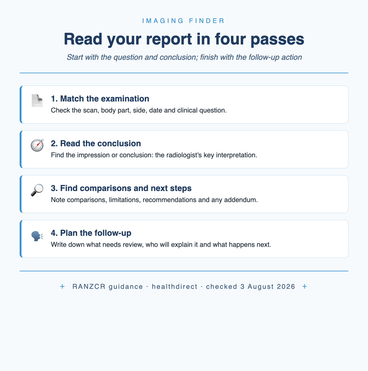
Short answer: read a radiology report in four passes. First, check the examination and clinical question. Second, read the impression or conclusion for the radiologist’s key interpretation. Third, find comparisons, limitations, recommendations and any addendum. Fourth, write down what needs follow-up and who will explain it. RANZCR says the conclusion should be concise and clinically relevant. Discuss the report in your clinical context rather than treating it as a stand-alone diagnosis or treatment plan.
On this page
What should you read first in a radiology report?
Begin above the medical detail. Check your name, the examination date, the scan type, the body part and the side. If the report says left knee and you expected a right-knee MRI, do not rely on it as your result: ask the imaging clinic or requester to confirm that the report belongs to your examination. RANZCR’s written-report guidance expects the report to identify the patient, examination and laterality where applicable.
Next, read the clinical details or indication. This is the question sent to the radiologist: for example, whether there is a fracture, a tendon tear or a cause for a particular symptom. The report should address the referrer’s specific clinical question, or explain why the scan could not answer it and what might answer it better.
Then go to the impression or conclusion. That section compresses the detailed observations into the radiologist’s clinically relevant interpretation. RANZCR also says normal or likely non-significant findings should be stated explicitly. A short X-ray report may not use a separate “Conclusion” heading, but the radiologist’s interpretation must still be part of the report.
Before reading the detailed Findings line by line, check the Comparison, any stated limitation, any recommendation and any addendum. Then work back through the Findings to see how the observations support the conclusion.
What does each section of the report mean?
Report layouts differ between hospitals, practices and scan types, but the jobs performed by the sections are recognisable. Some reports combine sections or use “Impression”, “Conclusion” and “Opinion” for the same final interpretive step.
A limitation describes something that reduced what the examination could show, such as movement or incomplete coverage; it is not itself a diagnosis. RANZCR says the report should name the limitation and explain how it affected interpretation.
Comparison deserves special attention. Current RANZCR standards require relevant prior studies to be compared when they are available. “Stable” or “unchanged” therefore means unchanged from the named earlier examination; it does not mean that the finding is absent.
If your earlier imaging was performed elsewhere and the Comparison section says none was available, bring that point to the follow-up appointment. The imaging appointment checklist explains how to keep reports and prior images together when you move between clinics.
What do uncertain words and recommendations mean?
Radiology is sometimes able to name a diagnosis from the images and sometimes only able to narrow the possibilities. RANZCR says non-specific or indeterminate findings should be labelled as such, with consideration given to how a more specific diagnosis might be reached. “Indeterminate” therefore means the imaging appearance is not specific enough for one answer. It does not, by itself, mean cancer or a harmless finding.
Words such as “likely”, “favoured” and “less likely” express the radiologist’s relative confidence. They are not promises. RANZCR advises radiologists to use commonly understood likelihood language and avoid vague phrases, so read the full conclusion rather than treating one qualifier as a verdict.
“Unremarkable” is usually the reassuring word it appears to be: no noteworthy abnormality was described for that structure on that examination. It is not a claim that every possible medical condition has been excluded. The same RANZCR guidance requires normal or likely non-significant findings to be explicit and any examination limitation to be explained.
Indeterminate is a decision point, not a diagnosis: read the sentence that follows. A recommendation might be comparison with older imaging, another test, clinical review or follow-up after a stated interval.
A recommendation should be more than “consider another scan”. RANZCR says it should be precise and explain how the proposed imaging, investigation, referral or treatment contributes to diagnosis or management. Your useful question is not “Is this bad?” but “What problem is the recommendation trying to resolve, and what timing applies to me?”
What should you do about an urgent finding or an addendum?
A report can be marked preliminary, provisional or final. A preliminary report may not yet have been checked for accuracy, content or typographical errors, and the final report can contain significant changes. For a routine review, check that the version you discuss is final. If a healthcare professional contacts you about an urgent result, follow that advice without waiting for a portal label to change.
An addendum is a clearly distinguished and dated addition or correction to the original report. When a later review differs in a way that could alter diagnosis or management, RANZCR says the people involved in care should be notified and the change documented with its author, date and time. Read the addendum together with the original impression rather than as a separate result.
Urgent and significant unexpected findings have a separate communication pathway. RANZCR requires the result to reach someone able to understand and act on it in a clinically appropriate timeframe, with escalation and a closed-loop process if the original referrer cannot be reached. The communication should also be documented.
That safety system does not turn silence into a normal result. Keep the result-review plan you made with the requester. If the report appears in your portal first and contains a recommendation or wording you do not understand, contact the healthcare professional who requested the scan and ask how the result changes the next step.
When can you see an imaging report in My Health Record?
The Australian Digital Health Agency says that from 1 July 2026, reports authored by or on behalf of a radiologist must be uploaded to My Health Record by default unless an exception applies or an extension has been granted. To use the patient-choice exception, you or your representative can tell the requesting clinician not to upload a particular report. If it has already been uploaded, you can remove it or control access in My Health Record. Other exceptions include a serious health or safety concern or circumstances outside the clinic’s reasonable control.
The display timing depends on the examination. The Australian Digital Health Agency says most limb X-ray reports are visible as soon as they are uploaded. Most other X-rays and reports for CT, MRI, PET, mammography and ultrasound become visible five days after upload. The five-day delay applies regardless of the result.
The default-sharing obligation covers the written report, not diagnostic images. My Health Record supplements existing information sources; the report is still sent directly to the requesting GP or clinician.
Inside My Health Record, select Documents, then Clinical Records and Diagnostic Imaging Reports. From that list, you can view or print a report, control access to it or remove it from your record.
What should you ask at the follow-up appointment?
Healthdirect describes the radiologist as the specialist doctor who interprets the images and summarises the result for your doctor. InsideRadiology advises discussing the report with the referring doctor so its meaning can be explained in your situation.
Take these questions to the result review
✓ “What question did this scan answer?”
✓ “Do any findings plausibly relate to my symptoms, and which findings are incidental?”
✓ “Was the relevant earlier imaging available for comparison?”
✓ “Does the recommendation apply to me, and what timing should we use?”
✓ “What should I do if my symptoms change before the next step?”
If the reviewing clinician does not already have them, bring the final report, the request and details of relevant earlier imaging. RANZCR identifies the clinical question and relevant prior comparison as core parts of interpretation. If the result leads to another scan, the diagnostic imaging request guide explains the written-request pathway, while the ultrasound preparation guide covers common scan-day instructions.
For background before that conversation, read the Medicare imaging overview and, if MRI was recommended, the MRI cost explainer.
If the conclusion does not account for the clinical problem, that does not automatically mean the scan was wrong. The examination may have a stated limitation, or the images may not show a cause for the symptoms supplied on the request. Discuss with the reviewing clinician whether observation, clinical review or another test is appropriate in your circumstances. The MRI-versus-CT guide explains why different examinations answer different questions.
What are common questions about radiology reports?
Which part of a radiology report is most important?
Start with the impression or conclusion because it contains the radiologist’s concise interpretation of the detailed observations. Then read the clinical question, comparison and any recommendation. A short report may not have a separately labelled conclusion, but RANZCR says the interpretation must still be present.
Does “unremarkable” mean normal?
It usually means no noteworthy abnormality was described for that structure on that examination. That is reassuring, but it is not a promise that every possible condition has been excluded. Read it with the examination type, any stated limitation and the conclusion.
Does “indeterminate” mean cancer?
No. It means the imaging appearance is not specific enough to give one diagnosis. RANZCR says the report should consider how a more specific diagnosis could be reached, such as comparison, follow-up or another test.
Why does the report mention an old scan?
Comparison shows whether a finding is new, unchanged or progressing. Current RANZCR standards require relevant prior studies to be compared when available. Check the named date so you know which earlier examination was used.
When will my imaging report appear in My Health Record?
From 1 July 2026, most limb X-ray reports are visible as soon as they are uploaded. Most other X-ray, CT, MRI, PET, mammography and ultrasound reports are visible five days after upload. An exception or clinic extension can mean a report is not uploaded; the report still goes directly to the requesting clinician.
Sources and methodology
The report structure and communication rules were checked on 3 August 2026 against current RANZCR standards, its written-report guideline and its critical-results position statement. Australian access requirements and timing come from the Australian Digital Health Agency. Healthdirect and InsideRadiology establish the patient-facing roles of the radiologist and referring doctor. These sources explain how reports communicate findings; they cannot interpret an individual report without the clinical context.
- RANZCR — Clinical Radiology Written Report Guidelines
- RANZCR — Standards of Practice for Clinical Radiology, version 12
- RANZCR — Clinical test result notification position statement
- Australian Digital Health Agency — Better access to health information
- Australian Digital Health Agency — diagnostic imaging reports
- healthdirect — radiographers and radiologists
- InsideRadiology — MRI information for adults
This article is general information, not personal medical advice. Discuss your individual report and next steps with the healthcare professional responsible for your care.

Author: Dr. Mark Bekhit
Dr Mark Bekhit (MBChB, FRANZCR) is a musculoskeletal radiologist practising in Brisbane and an AHPRA-registered specialist in diagnostic radiology (MED0002920947). He spends as much time building software as reading scans: he founded Imaging Finder (Australia) and Radiology Clinics NZ to make imaging easier to find, and builds radiology dictation and pain-tracking tools with one aim — better patient outcomes and a smoother reporting workflow. Outside the reading room he's usually training, bringing the same performance mindset to imaging.
Leave a Reply