By Dr. Mark Bekhit, Radiologist · Last updated August 2026
The quick answer
Short answer: For a scan your doctor has asked for, a CT is safe in the way that matters — the small added risk from the radiation is usually smaller than the risk of missing what the scan is looking for. A CT does use more radiation than a plain x-ray, but it adds to the natural background radiation everyone in Australia already receives every year. If you are or might be pregnant, tell the clinic. Ultrasound and MRI use none, and your doctor will use them where they fit.
On this page
- Why a CT uses more radiation than an x-ray
- Which scans use radiation, and which use none
- How much radiation is that, really?
- What actually matters: benefit vs risk
- Children and pregnancy
- How the clinic keeps your dose down
- Alternatives, and what to ask your doctor
- Find a CT clinic near you
- Common questions
- How we know: sources and method
Why a CT uses more radiation than an x-ray
A CT scan is a type of x-ray that builds detailed 2D or 3D pictures of the inside of your body. You lie flat and a ring-shaped machine turns around you; any part of the body can be scanned — the head, chest, abdomen or pelvis, or the heart in a coronary calcium score or CT coronary angiogram. The reason a doctor reaches for a CT rather than a plain x-ray is detail: it shows soft tissues, organs and blood vessels that do not show up well on an ordinary x-ray, which is what lets it diagnose an infection, check for injury after an accident, plan surgery or guide a biopsy.
That extra detail is exactly why a CT delivers more radiation. A plain x-ray is a single picture; a CT takes many x-ray images and combines them, so it uses a higher level of radiation and should only be used when it is genuinely needed. None of this radiation lingers: the radiation is only present while the machine is switched on, and having an x-ray or CT does not make you radioactive. Once the scan is over, there is nothing radioactive left in the room or in you.
Which scans use radiation, and which use none
Not all imaging uses radiation, and it helps to know where a CT sits. The x-ray family — plain x-rays, mammograms, fluoroscopy and CT — all use ionising radiation. Ultrasound and MRI do not use or produce any ionising radiation at all — an MRI works with strong magnets and radio waves instead of x-ray radiation. Among the x-ray tests, a plain x-ray uses only a small amount of radiation, and plain x-rays and mammograms give a lower dose than a CT.
This is the first thing a radiologist thinks about when a request comes in. If ultrasound or MRI can answer the question, they are usually preferred — not because a CT is unsafe, but because there is no reason to use radiation when a test without it will do the same job.
How much radiation is that, really?
Numbers help here, and the honest ones are more reassuring than the scary figures that circulate online. Radiation dose is measured in millisieverts (mSv), a unit that combines the dose and its likely risk. The useful yardstick is the radiation you are already getting for free from the world around you. In Australia, natural background radiation is about 2 mSv a year — roughly the equivalent of 100 chest x-rays — from cosmic rays, the ground, building materials, and the food we eat. Everyone in Australia soaks that up every year, simply by living here.
A CT sits above a plain x-ray but is still measured against that same everyday backdrop, not against some special category of danger. Beware pages — often from overseas — that quote a different background figure or an individual “chance of cancer” from one scan. Those numbers are drawn from other countries and, as below, cannot honestly be applied to you as a person.
What actually matters: benefit vs risk
“Is a CT safe?” is really two questions. Does it carry any risk? A little. Is that risk worth it? Almost always, when a doctor has requested the scan. Exposure to radiation above background slightly increases the risk of developing cancer, but the increase is small — and usually less than the risk of not identifying or treating a problem properly. A CT that finds a treatable cancer early, or rules out bleeding after a fall, usually protects you far more than the small dose is likely to harm you.
It is worth being clear about what that risk is not. The long-term risk from medical radiation is so small it is expressed as chances per million, and it cannot be calculated for any one person — the estimates are averages across large populations. So when a website offers you a personal “your CT gives you an X% chance of cancer” figure, it is overstating what the science can actually say. The genuinely useful rule is the one radiologists use: every scan should have a definite benefit to balance its small risk. If you are unsure a scan clears that bar, that is the conversation to have with your referring doctor — not a reason to quietly skip it.
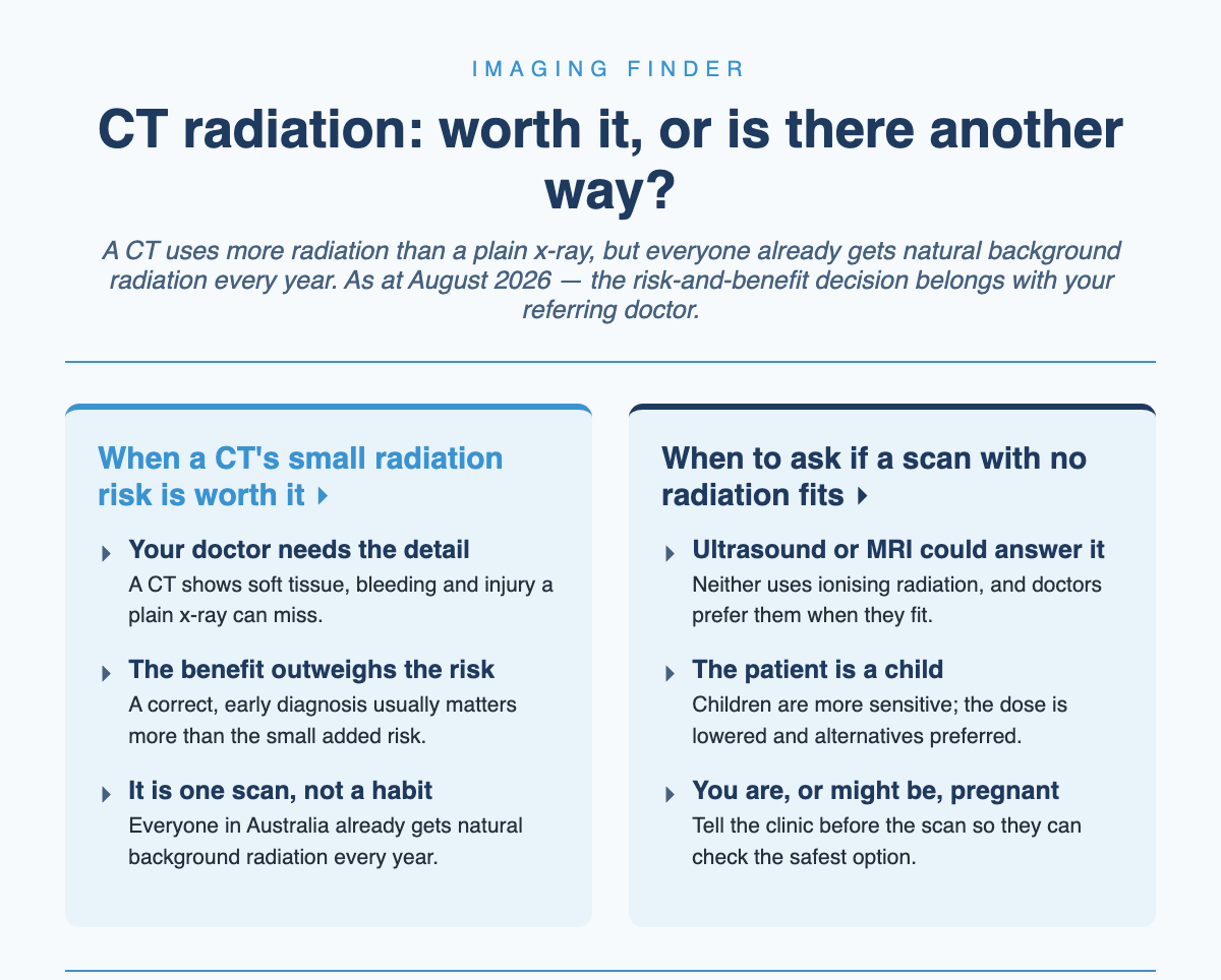
The one question worth asking your doctor. Not “how much radiation?” but “what would we do if I didn’t have this scan?” If the answer is that your doctor needs the pictures to diagnose or treat you safely, the benefit almost certainly outweighs the small radiation risk. If a test without radiation would answer the same question, your doctor can weigh that too — and the radiologist supervising the scan also checks it is the most appropriate test for you.
Children and pregnancy
Two groups deserve extra care, and the sources are clear about both. Children are more sensitive to radiation than adults — their tissues are growing and dividing faster, and they have more years ahead for any effect to appear. That does not mean children cannot have a CT; it means the dose is handled carefully. A child’s dose is adjusted for their smaller body size, a modern scanner is used, and where a test without radiation — ultrasound or MRI — can answer the question, it is preferred.
In pregnancy, the instinct to worry is natural, and the reassurance is real — and if you want the whole picture across scan types, see our guide to whether medical imaging is safe during pregnancy. A CT is generally best avoided in pregnancy where another test will do, so tell your doctor and the clinic if you are pregnant or think you might be. But if a CT is genuinely needed, the numbers are on your side: international radiation-protection guidance sets a threshold of 100 mGy below which harm to a developing baby is not expected, and even the highest-dose examinations give a single-scan dose to the baby that is well below that threshold. The decision is one to make with your doctor, but it is not the catastrophe it can feel like.
How the clinic keeps your dose down
A lot of safety is built into the scan before you even arrive, and it is fair to expect it. Radiographers are trained to use the lowest dose that still gives clear pictures, they scan only the part of the body that needs it, and they may use lead or bismuth shields to protect sensitive areas. The equipment itself is maintained and calibrated by specialists under State and Federal law, and the people operating it must be trained and licensed. This principle — use only as much radiation as the job needs — is the industry’s standard, not an optional extra.
One point of scope: this article is about the x-ray radiation of a CT. If your CT uses an injected contrast dye, that is a separate consideration with its own small risks, which I cover in the guide to CT contrast dye safety.
Alternatives, and what to ask your doctor
Sometimes a scan without radiation fits the question just as well, and it is reasonable to ask. MRI and ultrasound are used in preference to other tests whenever they can answer what the doctor needs, and your referring doctor chooses the most appropriate test for your situation — and can talk it through with the radiologist. Our guide to whether you need an MRI or a CT walks through when each one is the better tool.
Before your CT, it is worth doing this:
- Tell the clinic if you are pregnant or think you might be, so they can check the safest option.
- Ask your referring doctor what the scan will show and how it will change your care — the benefit is the point.
- Ask whether a test without radiation, like ultrasound or MRI, could answer the same question.
- For a child, ask that the dose be adjusted for their size on a modern scanner.
- Bring any previous scans or reports so the team is not repeating imaging you have already had.
Find a CT clinic near you
When your doctor has decided a CT is the right test, the next step is booking it somewhere convenient. You can search CT clinics near you on Imaging Finder — the directory lists CT clinics across the metros, including Melbourne, Brisbane and Perth, so you can compare locations and call ahead. When you book, it is also a good moment to check what a scan will cost; our guide to CT scan costs in Australia explains the Medicare rebate and gaps, and the referral guide covers who can request one.
Common questions
Is one CT scan dangerous?
No. A single CT that your doctor has requested carries only a small added risk, which is usually smaller than the risk of leaving a problem undiagnosed. The dose adds to the background radiation everyone already receives every year, and the benefit of the diagnosis is the reason the scan is worth having.
How much radiation is in a CT scan?
A CT uses more radiation than a plain x-ray because it combines many images. The exact amount depends on the part of the body and the scanner, so rather than a single figure it is most honest to say a CT is a meaningful step up from a plain x-ray, measured against the roughly 2 mSv of natural background radiation Australians receive each year. Your radiology clinic can tell you more for your specific scan.
Can I have a CT scan while pregnant?
Sometimes, if it is genuinely needed — but a CT is best avoided in pregnancy where another test will do, so tell your doctor and the clinic. If a CT is necessary, the dose to a baby from a single scan is well below the level at which harm would be expected. Make the decision with your doctor.
Does a CT scan make me radioactive?
No. The radiation is only present while the machine is on, and none remains in your body or the room afterwards. You are safe to be around children and pregnant people straight after a CT. (Nuclear medicine scans, which use an injected radioactive tracer, are the exception — they make you slightly radioactive for a short time, but a CT does not.)
Is an MRI or ultrasound safer than a CT?
Neither uses any ionising radiation, so where one of them can answer your doctor’s question it is usually preferred. But they are not interchangeable — a CT is sometimes the only test that shows what is needed. The right choice depends on the clinical question, which is why your doctor and the radiologist decide it together.
How we know: sources and method
Every decisive figure in this article is drawn from current Australian sources, read on 22 August 2026: healthdirect (the Australian Government health-information service) on what a CT is and its radiation; and InsideRadiology, the patient-information service of the Royal Australian and New Zealand College of Radiologists (RANZCR), on the radiation risk of medical imaging for adults and children, radiation risk during pregnancy, and how a CT is carried out. Radiation doses are given against the Australian background figure rather than overseas numbers, and no personal cancer-risk figure is quoted because, as the sources explain, such a figure cannot be calculated for an individual.
This article is general information, not personal medical advice. Discuss your individual report and next steps with the healthcare professional responsible for your care.

Author: Dr. Mark Bekhit
Dr Mark Bekhit (MBChB, FRANZCR) is a musculoskeletal radiologist practising in Brisbane and an AHPRA-registered specialist in diagnostic radiology (MED0002920947). He spends as much time building software as reading scans: he founded Imaging Finder (Australia) and Radiology Clinics NZ to make imaging easier to find, and builds radiology dictation and pain-tracking tools with one aim — better patient outcomes and a smoother reporting workflow. Outside the reading room he's usually training, bringing the same performance mindset to imaging.
Leave a Reply